Schedule
Workshop Date: October 8th 2020, Provisional Programme.
14:00 - 14:10UTC Introduction
14:10 - 15:00UTC Virtual Keynote by Polina Golland, followed by live Q/A session.
15:00 - 16:00UTC Online Oral Session 1, with live Q/A session.
15:00-15:10: Deformable Slice-to-Volume Registration for Reconstruction of Quantitative T2* Placental and Fetal MRI, Alena Uus
15:10-15:20: Harmonised segmentation of neonatal brain MRI: a domain adaptation approach, Irina Grigorescu
15:20-15:30: Unbiased atlas construction for neonatal cortical surfaces via unsupervised learning, Lilla Zöllei
15:30-15:40: Brain volume and neuropsychological differences in extremely preterm adolescents, Hassna Irzan
15:40-15:50: 3D Fetal Pose Estimation with Adaptive Variance and Conditional Generative Adversarial Network, Junshen Xu
15:50-16:00: Live virtual Q&A Session with all the presenters of the session
16:00 - 17:00UTC Online Coffee with ePoster Drop-in Sessions and focused Speaker Discussions.
1. Multi-Modal Perceptual Adversarial Learning for Longitudinal Prediction of Infant MR Images, Liying Peng
2. Spontaneous preterm birth prediction using convolutional neural networks, Tomsz Wlodarczyk
3. Automatic Detection of Neonatal Brain Injury on MRI, Russel MacLeod
4. Efficient multi-class fetal brain segmentation in high resolution MRI reconstructions with noisy labels, Kelly Payette
5. Maternal irritability during pregnancy predicts decreased infant frontoparietal, Sanjana Ravi
6. Automatic Segmentation of the Placenta in BOLD MRI, Mazdak Abulnaga
7. Atypical Neonatal Extra-axial CSF is Associated with Reduced Cognitive Development at Age 1 year, Martin Styner
8. Fetal brain masking: evaluating the generalizability of an automated toolbox, Emily Nichols
9. In vivo tractography of the white-matter tracts associated with the reward network and hypothalamus in the neonatal brain, Julie Nihouarn
10. Regional brain volumes at 19 years of age following extremely preterm birth and the effect on intelligence, Ramya Thanikasalem
17:00 - 17:45UTC Virtual Invited Talk by Michael Aertsen, followed by live Q/A session.
18:00 - 19:00UTC Online Oral Session 2, with live Q/A session.
18:00-18:10: Deep Learning Spatial Compounding from Multiple Fetal Head Ultrasound Acquisitions, Jorge Perez-Gonzalesz
18:10-18:20: Automated Detection of Congenital Heart Disease in Fetal Ultrasound Screening, Jeremy Tan
18:20-18:30: Multi-task approach using positional information for ultrasound placenta segmentation, Veronika Zimmer
18:30-18:40: Atlas-based segmentation of the human embryo using deep learning with minimal supervision, Wietske Bastiaansen
18:40-18:50: A Smartphone-based System for Real-time Early Childhood Caries Diagnosis, Yipeng Zhang
18:50-19:00: Live virtual Q&A Session with all the presenters of the session
19:00 - 19:15UTC Open Discussion, Best Paper Award and Closing.
PIPPI 2020 Keynote Talk
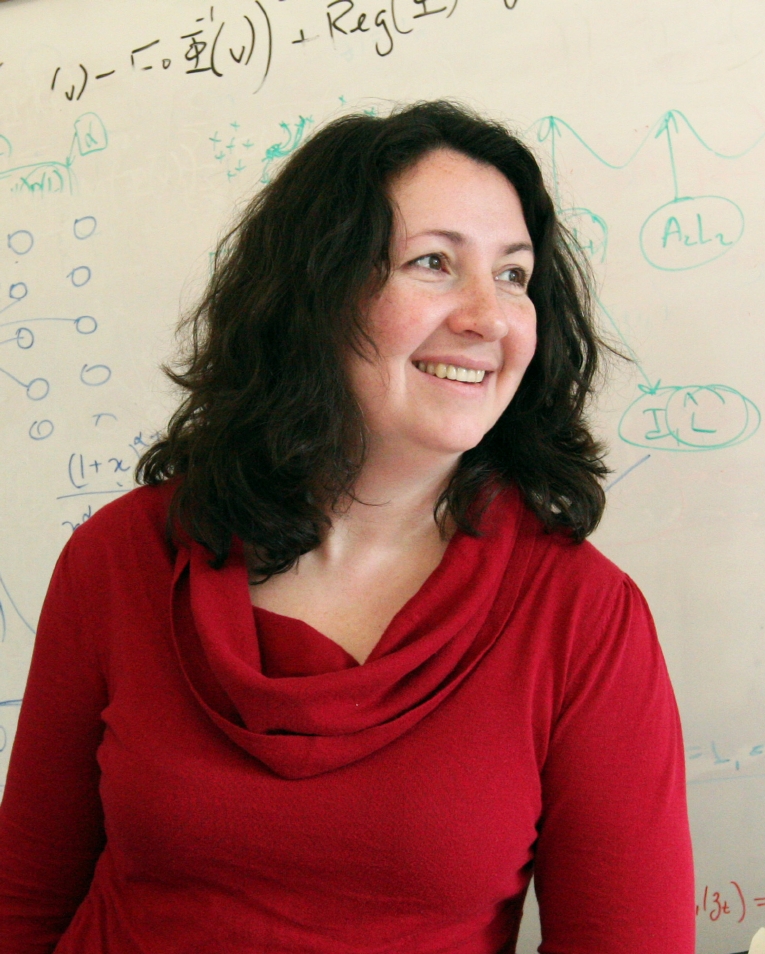 Polina Golland
From Pixels to Clinical Insight: Placental MRI Analysis
Placental MRI presents many challenges for constructing biomarkers of
normal development and pathology. We introduce a set of computational
tools that support image based studies of the placental anatomy and
function. The goal of the talk is to encourage discussion of open
problems and potential directions for advancing our understanding of
the placenta through imaging and analysis.
Polina Golland is a Henry Ellis Warren (1894) professor of Electrical
Engineering and Computer Science at MIT and a principal investigator
in the MIT Computer Science and Artificial Intelligence Laboratory
(CSAIL). Her primary research interest is in developing novel
techniques for medical image analysis and understanding. With her
students, Polina has demonstrated novel approaches to image
segmentation, shape analysis, functional image analysis and population
studies. She has served as an associate editor of the IEEE
Transactions on Medical Imaging and of the IEEE Transactions on
Pattern Analysis. Polina is currently on the editorial board of the
Journal of Medical Image Analysis. She is a Fellow of the
International Society for Medical Image Computing and Computer
Assisted Interventions.
PIPPI 2020 Invited Talk
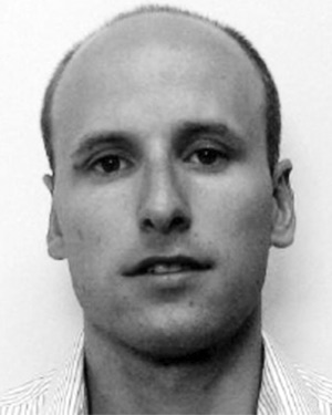 Michael Aertsen
Fetal MRI of the central nervous system
Fetal MRI is an important adjunct to prenatal ultrasound for the detection exploration and prognostication of in utero abnormalities. In the beginning maternal sedation was used but this was abandoned due to improved acquisition techniques. Furthermore there is an increasing use of more functional techniques (DWI, DTI, BOLD imaging) to add in the prognostication of certain fetal abnormalities. With the growing applications of AI, new possibilities are available in fetal imaging due to motion correction allowing for volumetric analysis, shape analysis and more robust functional imaging.
Michael Aertsen started a part-time Phd project on ‘The role of advanced fetal imaging in current fetal medicine’ in 2015 after being involved in fetal MR at the University Hospitals for 2 years. From 2015 he was involved in all prenatal and post mortem studies concerning fetal MR in University Hospitals Leuven and regarding artificial intelligence developed at University College London. In 2017 he conducted a retrospective analysis of the posterior fossa alterations in spina bifida aperta. In addition, he also developed a low dose fetal CT protocol for severe skeletal abnormalities. In 2018 he launched the postmortem micro CT to evaluated small fetuses (18 weeks of gestation) and investigate fetal hearts in congenital heart defects as well as normal comparisons. In 2019 he started a large prospective study to examine the normal fetal brain on 3 Tesla MR with anatomic, diffusion weighted and spectroscopy.
He is member of the editorial board of European Journal of Radiology and Prenatal Diagnosis.