MICCAI Workshop on Perinatal, Preterm and Paediatric Image analysis (PIPPI)
PIPPI was successfully held on Friday 21st October 2016. For more information, please contact the workshop organisers
Objectives
The application of sophisticated analysis tools to postnatal, infant and paediatric imaging data is of interest to a substantial proportion of the MICCAI community. The field has grown substantially since the most recent perinatal themed MICCAI satellite workshop in 2012. The main objective of this workshop is to bring together researchers in the MICCAI community to discuss the challenges of image analysis techniques as applied to the preterm, perinatal and paediatric setting.
Advanced medical image analysis allows the detailed scientific study of conditions such as prematurity and the study of both normal singleton and twin development in addition to less common conditions unique to childhood. This workshop will bring together methods and experience from researchers and authors working on these younger cohorts and will provide a forum for the open discussion of advanced image analysis approaches focused on the analysis of growth and development in the perinatal, preterm and paediatric period.
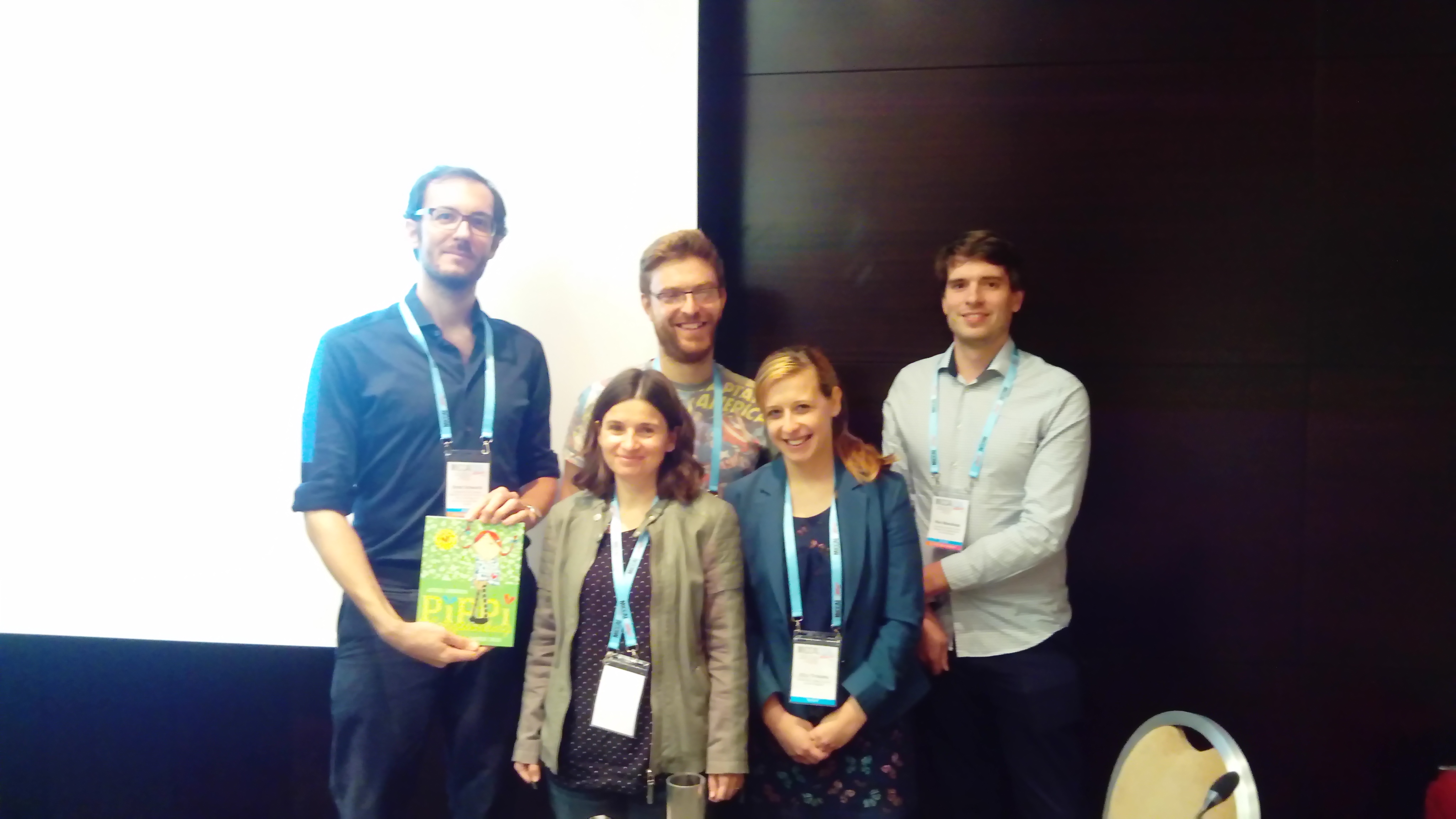
Best Paper Prize 2016
Keynote Lecture
An overview of the developing human connectome project (DHCP)
Daniel Rueckert
Imperial College London

Few advances in neuroscience could have as much impact as a precise global description of human brain connectivity (connectome) and its variability. Understanding this connectome in detail will provide insights into fundamental neural processes and intractable neuropsychiatric diseases. Currently, the connectome of the mature adult brain is in progress. The Developing Human Connectome Project (dHCP) aims to make major scientific progress by creating the first 4-dimensional connectome of early life. Our goal is to create a dynamic map of human brain connectivity from 20 to 44 weeks post-conceptional age, which will link together imaging, clinical, behavioural, and genetic information. This unique setting, with imaging and collateral data in an expandable open-source informatics structure, will permit wide use by the scientific community, and to undertake pioneer studies into normal and abnormal development by studying well-phenotyped and genotyped group of infants with specific genetic and environmental risks that could lead to Autistic Spectrum Disorder or Cerebral Palsy.
Topics of interest
Potential methods will cover the full scope of medical image analysis, but there must be an application to younger cohorts or to the long term outcomes of perinatal conditions.
Topics may include:
- Image Registration, Segmentation or Classification
- Image Reconstruction
- Atlas Construction
- Diffusion Imaging (including tractography applications)
- Longitudinal and Cross-sectional Studies
- Advanced structural imaging (e.g. advanced DWI or g-ratio analysis)
- Advanced functional imaging (e.g. network-based analysis)
- Fetal and/or placental image analysis
- Cardio-pulmonary Image analysis
- Abdominal Imaging
- Measurement of long-term cognitive outcome
- Correlation of imaging biomarkers with functional measurements
Important Information
Workshop Date: October 21st (AM)
Workshop Location: Intercontinental Athenaeum, Athenaeum CCII
Why Submit to PIPPI?
- To disseminate new work to the most relevant members of the MICCAI community
- To take part in discussion of new techniques for imaging of younger cohorts.